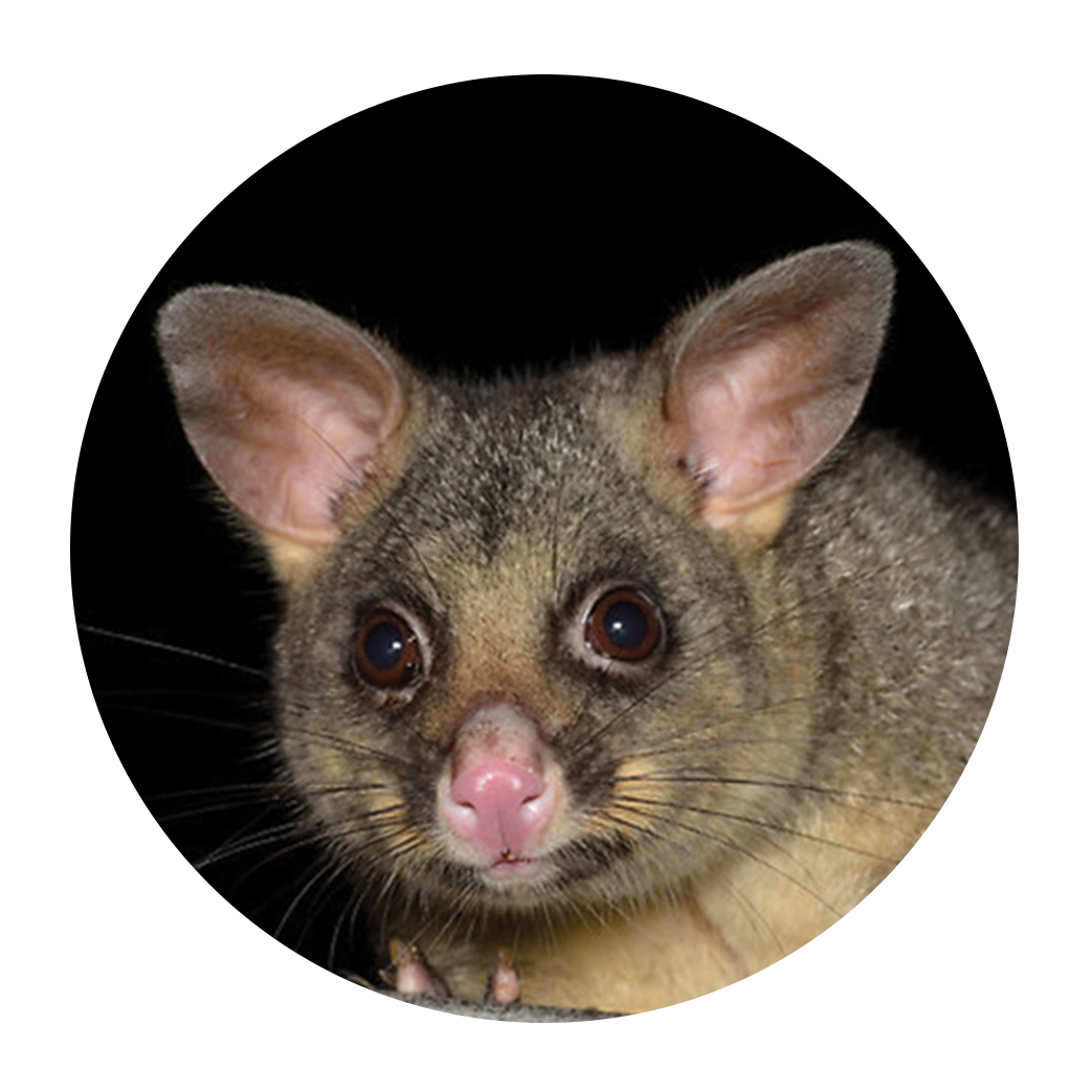
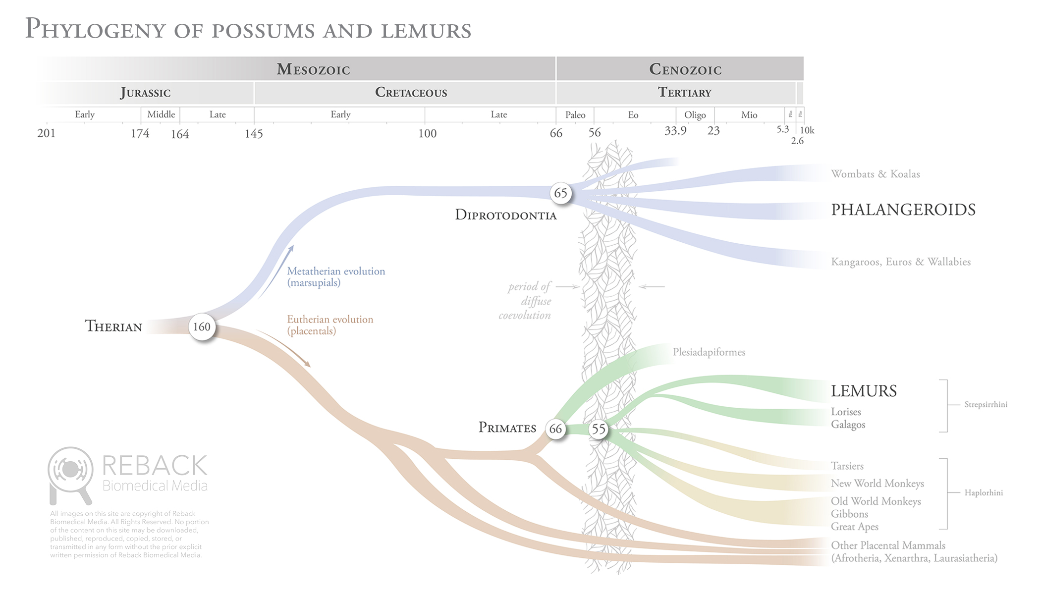
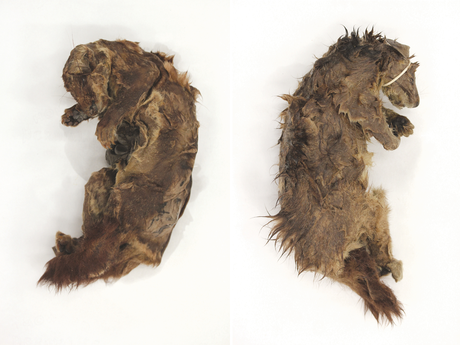
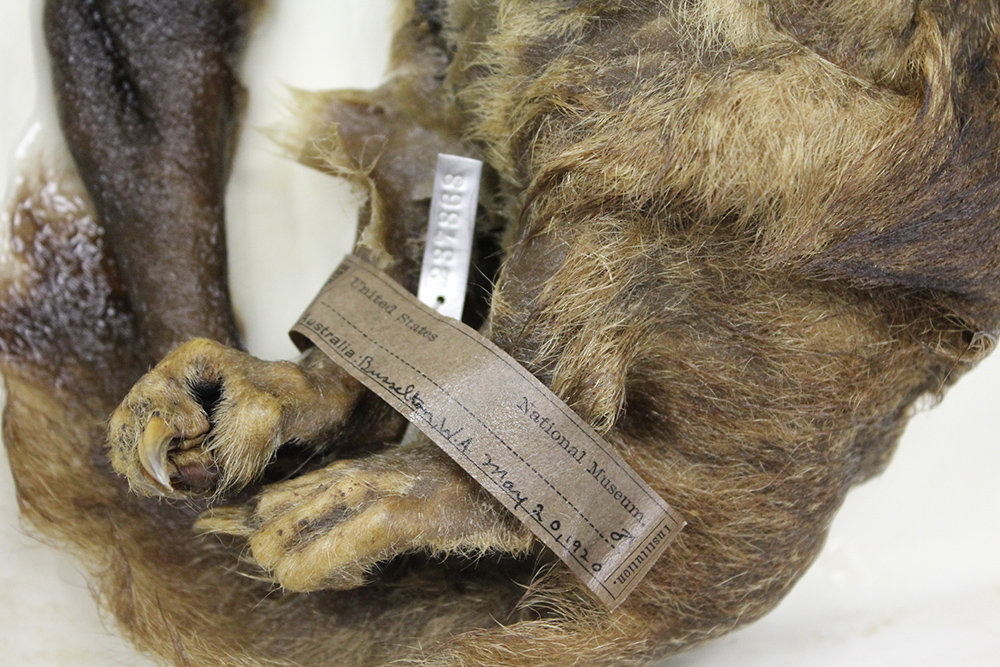
JHU – Master’s Thesis

Masters Thesis

An educational three dimensional model to describe the masticatory apparatus of the phalangeroid possum, Trichosurus vulpecula

Developed in collaboration with the following institutions at the Johns Hopkins University:

purpose

No mammal can survive without eating. Understanding the morphology of the masticatory apparatus tells us what foods a mammal is best adapted to consuming be it plant, animal, or both. It also allows us to understand how a mammals ecological niche may have influenced the evolution of its masticatory apparatus and other structures.

The primary goal of this project was to digitally reconstruct the masticatory apparatus of a single species of phalangeroid possum, Trichosurus vulpecula. The reconstruction serves as a visual communication tool that improves the ability of the evolutionary biologist to make meaningful comparisons between the phalangeroid possums of New Guinea and Australia to lemurs of Madagascar. Lemurs, a diverse group of primitive primates, are important in the study of primate origins.

The secondary goal of this project was to document the digital reconstruction workflow to serve as an instructive guide for those interested in making their own 3D representations of mammalian masticatory apparatuses.

Why study Phalangeroids?

The phalangeroid possums of New Guinea and Australia are a group of mammals possessing many morphological and biological traits that are highly similar to the lemurs of Madagascar. Lemurs and phalangeroids are an example of a phenomenon known as convergent evolution: they are not closely related, yet developed similar traits as a result of having to adapt to similar ecological niches. 

PHALANGEROID POSSOM

Trichosurus vulpecula
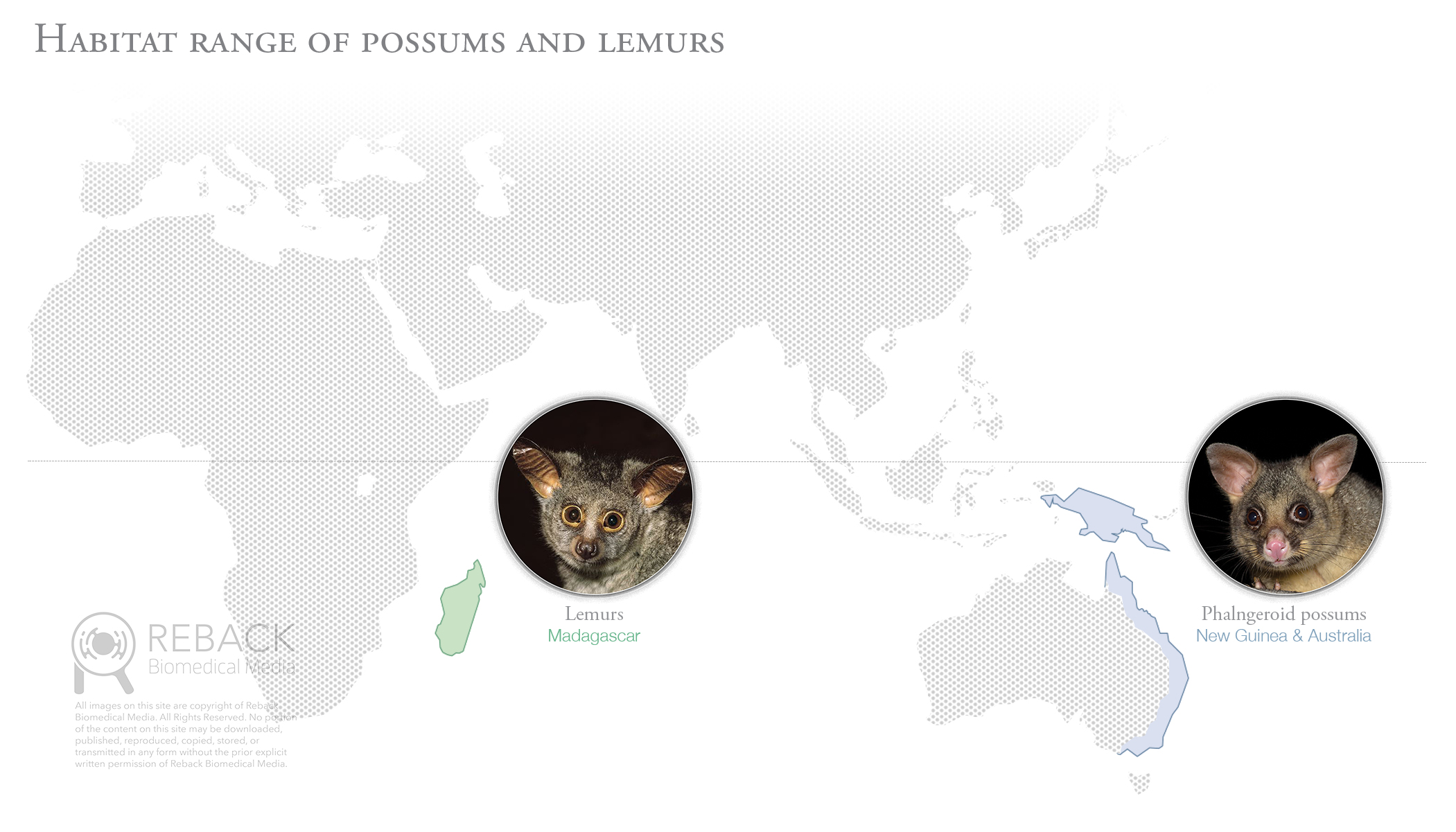
A marsupial mammal of the family Phalangeridae native to the island of New Guinea and east coast of Australia.
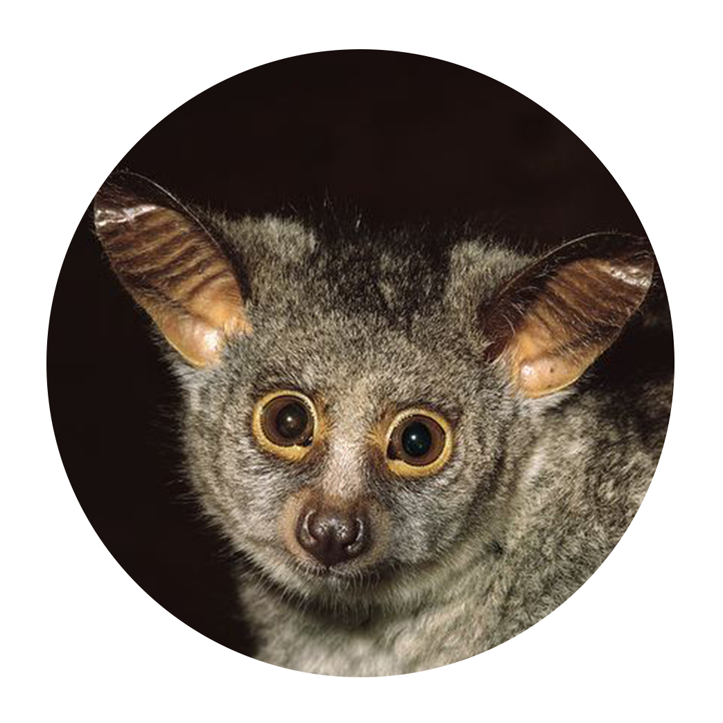
LEMUR

Otolemur crassicaudatus
A placental mammal of the order Primate native to the island of Madagascar.

Background

isolated habitats

Possums and lemurs evolved in separate and isolated regions of the world, however, the environmental conditions in each geographic region are highly similar.

Shared traits

The similarity of ecological niches inhabited by possums and lemurs influenced these two unrelated groups of mammals to develop highly similar traits.

Independent evolution

The last common ancestor to both possums and lemurs was a lizard-like mammal called a therian who lived approximately 160 mya. In fact, the diversity of all mammalian life can be traced back to this single predecessor.

reconSTruction process

Dissection of several preserved Trichosurus vulpecula specimens took place at the Smithsonian National Museum of Natural History Museum Support Center outside of Washington, D.C. Data was gathered on muscle mass, fiber direction, and areas of origin and insertion. 

results

anatomical landmarks

Illustration was used to document the anatomical landmarks of the cranium and mandible. Observations gathered during dissection helped determine locations of muscle origin (red) and insertion (blue).

3D reconstruction

Hard tissue reconstruction relied upon segmenting data acquired from a micro-CT scan of a Trichosurus skull. Muscles were created from scratch using digital sculpting software. The mass of each muscle was matched to the average weight taken from
dissected specimens.

3D Animation

A turntable animation was created to give the audience an overall sense of spatial relationships in the masticatory apparatus. An exploded view animation demonstrates the layering of the muscles. Deeper structures, such as the pterygoids, are visualized by seeing through the mandible.

Play Video

A new TOOL FOR VISUAL COMMUNICATION

Articulated 3D PRINTED MODEL

The final output of this project was an articulated 3D printed model at 200% scale. Neodymium magnets were used to allow the audience to take apart and reassemble the muscle layers as well as separate the mandible from the skull. An axis was created in the temporomandibular joint allowing the mandible to be articulated when the muscles are removed. Several prototypes were created to design and test various physical features that made the model easy and fun to interact with.

acknowledgements

I would like to thank my thesis advisor, Jennifer Fairman, MA, CMI, FAMI, and preceptor Jonathan M.G. Perry, MSc, PhD, for their intellectual, strategic, and emotional support throughout the project. I would also like to thank my classmates, particularly Sarah A. Chen, MA, MD, for her encouragement to step away now and then and have a beer (or two). My family and friends are a constant source of support in anything I do, so a special thanks goes out to them as well.

Click the button below to download a free pdf copy of the full thesis, including tips and techniques on how to create digital 3D reconstructions of your own.

Reback Biomedical Media | 608.852.3233 | rebackbiomed@gmail.com

All content © Reback Biomedical Media, 2015-2021, unless otherwise noted. All rights reserved.

JHU – Reno Epidural Anastomosis

Embolization of the Reno epidural anastomosis for nutcracker syndrome

PROJECT

Pathology 2D animation

AUDIENCE

Candidates for surgical treatment of Nutcracker syndromen

DESCRIPTION

Renal nutcracker syndrome is a rare disorder where blood flow coming out of the kidney is restricted. The restriction is caused by compression of the left renal vein by the superior mesenteric artery. In moderate to severe cases, patients can experience blood in the urine (hematuria) and chronic abdominal pain, pelvic pain, flank pain, fatigue, or a combination thereof.

The purpose of this animation is to educate patients on the anatomy of nutcracker syndrome and provide and overview of several surgical treatments. One such treatment involves placing a stent within the left renal vein at a site beneath the superior mesenteric artery to prevent compression. Another treatment involves embolization of the left gonadal and/or renal epidural anastomosis to raise pressure within the left renal vein and prevent collapse.

Reback Biomedical Media | 608.852.3233 | rebackbiomed@gmail.com

All content © Reback Biomedical Media, 2015-2021, unless otherwise noted. All rights reserved.

JHU – Biological Filtration

Aerobic biological filtration for freshwater aquariums

PROJECT

Biological illustration

AUDIENCE

Aquarium technicians and home enthusiasts

DESCRIPTION

Ammonia (NH3) is a waste product excreted by fish and the decomposition of uneaten food. NH3 is toxic to fish and must be continuously filtered out of a closed system like an aquarium. Fortunately, microorganisms called nitrifying bacteria can consume NH3 as their source of energy, eventually converting it to a non-toxic substance called nitrate (NO3).

This set of illustrations depicts three different designs for aerobic biological filtration of nitrogenous waste in aquariums. Water passes through a high surface area medium (ie: gravel, bio ball, or sponge) that permits colonization of nitrifying bacteria. Oxygen is diffused into the water by either agitation (biotower filter) or pumping room air through an air stone (under gravel or sponge filter).

Details

INSETS

For more information on the nitrogen cycle in aquariums,
check out the online resources listed below:

Florida Department of Agriculture and Consumer Services:
Aquarium water quality: Nitrogen cycle

The aquarium co-op:
The easy guide to the nitrogen cycle for aquariums

Fishlore:
Aquarium nitrogen cycle – new tank syndrome


Reback Biomedical Media | 608.852.3233 | rebackbiomed@gmail.com

All content © Reback Biomedical Media, 2015-2021, unless otherwise noted. All rights reserved.

JHU – Surgical Instruments

3D modeling of surgical instruments

PROJECT

Cinema 4D three dimensional modeling and photorealistic rendering

AUDIENCE

General medical audience

DESCRIPTION

The purpose of this project was to develop basic 3D modeling capabilities in Cinema 4D for hard surface objects. A small variety of ubiquitous surgical instruments were chosen as subjects for the assignment. Importance was placed on dimensional accuracy, mechanical details, touch points, as well as color, material, texture and graphics.

Instruments were placed in a simulated studio environment with a two-light set up. Photorealistic renderings were created using the physical renderer with ambient occlusion and an HDRI studio rig to increase the perception of realistic reflections.

Reback Biomedical Media | 608.852.3233 | rebackbiomed@gmail.com

All content © Reback Biomedical Media, 2015-2021, unless otherwise noted. All rights reserved.

JHU – Carotid Body Tumor

Surgical Removal of
a Carotid Body Tumor

PROJECT

Surgical illustration 

AUDIENCE

Vascular and head/neck surgeons

DESCRIPTION

A carotid body tumor (also known as a chemodectoma or paraganglioma) is a rare, highly vascular mass that forms within the carotid sheath at the base of the carotid bifurcation. Tumors are often benign but may cause impairment of adjacent cranial nerves resulting in loss of associated function.

The purpose of this illustration is to teach the main steps of the surgery with special attention paid to the reaction of tissue to the instruments and other forces applied to it. The illustration was produced in collaboration with surgeons in the department of otolaryngology at the Johns Hopkins Hospital in Baltimore, Maryland. 

INSETS

layout For a surgical atlas

Reback Biomedical Media | 608.852.3233 | rebackbiomed@gmail.com

All content © Reback Biomedical Media, 2015-2021, unless otherwise noted. All rights reserved.

JHU – Quadrangular Space

Innervation in the quadrangular and triangular space of the shoulder

PROJECT

Anatomical plate

AUDIENCE

Educators and students of human anatomy

DESCRIPTION

The spatial relationships of the posterior shoulder are complex and difficult to visualize. Nerves course between openings created by the boundaries of several overlapping muscles.

The purpose of this illustration is to introduce innovative storytelling techniques as a means to improve upon previous depictions of a challenging aspect of human anatomy. A few of the techniques used in this illustration are: 1) Anatomy shown in context of the figure; 2) Instructional color to visually organize innervation and correlate specific nerves with their anatomical windows of origin; 3) Metaphor to help the audience form an abstract mental image of key spatial relationships.

Details

Reback Biomedical Media | 608.852.3233 | rebackbiomed@gmail.com

All content © Reback Biomedical Media, 2015-2021, unless otherwise noted. All rights reserved.

JAMA – Medical News

Medical news & perspectives

Articles in the Medical News & Perspectives section are focused on current events in healthcare and medical science. In-house writers chose a topic of interest and a medical illustrator is assigned to capture a particular aspect of the story in an editorial-style illustration. 

In contrast to clinically focused illustrations, those produced for Medical News are emotive and often experimental in style and technique. Although each illustration aims to be instructive in nature, the tone varies widely between them.

ARTICLE LINKS

COPYRIGHT NOTICE

All illustrations displayed on this post are copyright of the American Medical Association. All rights reserved.

Reback Biomedical Media | 608.852.3233 | rebackbiomed@gmail.com

All content © Reback Biomedical Media, 2015-2021, unless otherwise noted. All rights reserved.

JAMA – Patient Pages

PATIENT INFORMATION

JAMA Patient Information pages are a free source of one page easy-to-understand articles on prevention and management of illness or other conditions. Each article is authored by a clinician working in their specialty with whom a JAMA medical illustrator works to develop an accompanying illustration that helps explain the topic to a non-medical audience.

Access to Patient Information pages is free. No membership to the American Medical Association is required. Use the links below to view articles for each illustration shown here, or search for another topic using the JAMA Patient Information web portal.

ARTICLE LINKS

1. Patellofemoral pain
01.23.18 – JAMA

3. Vasectomy
06.19.18 – JAMA

4. Gallbladder cancer
09.25.18 – JAMA

6. Focal radiation therapy for cancer
01.10.19 – JAMA Oncology

8. Sigmoid volvulus
06.25.19 – JAMA

11. What is Mohs surgery?
05.06.20 – JAMA Dermatology

13. What are ear tubes?
10.15.20 – JAMA Otolaryngology

14. Bladder cancer
11.17.20 – JAMA

15. Urinary diversion
TBD – JAMA

COPYRIGHT NOTICE

All illustrations displayed on this post are copyright of the American Medical Association. All rights reserved.

Reback Biomedical Media | 608.852.3233 | rebackbiomed@gmail.com

All content © Reback Biomedical Media, 2015-2021, unless otherwise noted. All rights reserved.

JAMA – Difficult Intubation

WILL THIS PATIENT BE DIFFICULT
TO INTUBATE - THE RATIONAL
CLINICAL EXAMINATION

PUBLICATION DATE

02.05.2019

DESCRIPTION

Endotracheal intubation (ETI) is often employed during major surgical procedures and for respiratory support in patients who are critically ill. Not all patients can be intubated easily, however, and failure of advanced recognition of difficult intubation can result in a catastrophic scenario.

The purpose of this illustration is to demonstrate several anatomic features and bedside tests that can be used to assess difficult intubation prior to general anesthesia or in emergency situations. This set of illustrations is unique in that it combines the most common strategies for airway assessment within a single easy-to-understand composition.

ARTICLE LINK

AMA membership required for full article access.

COPYRIGHT NOTICE

All illustrations displayed on this post are copyright of the American Medical Association. All rights reserved.

Upper Lip Bite Test

Figures in context

Reback Biomedical Media | 608.852.3233 | rebackbiomed@gmail.com

All content © Reback Biomedical Media, 2015-2021, unless otherwise noted. All rights reserved.

JAMA – Pelvic Organ Prolapse

Pelvic Organ prolapse Quantification -
An original investigation

PUBLICATION DATE

04.17.2018

DESCRIPTION

Female pelvic organ prolapse (POP) involves the downward descent of pelvic and/or lower abdominal organs resulting in protrusion of the vagina, uterus, or both. The condition is becoming more common given the aging population in the United States.

The purpose of this illustration, using normal anatomy of a nulliparous female, is to demonstrate the precise location of anatomic points used to assess POP during examination. The illustration is unique in that it is the first to unequivocally document these points and provide precise examples POP stages.

ARTICLE LINK

AMA membership required for full article access.

COPYRIGHT NOTICE

All illustrations displayed on this post are copyright of the American Medical Association. All rights reserved.

INSETS

FIgure in Context

Reback Biomedical Media | 608.852.3233 | rebackbiomed@gmail.com

All content © Reback Biomedical Media, 2015-2021, unless otherwise noted. All rights reserved.

JAMA – OSA

Diagnosis and Management of Obstructive Sleep Apnea -
A review

PUBLICATION DATE

04.14.2020

DESCRIPTION

Obstructive sleep apnea (OSA) is characterized by recurrent episodes of partial or complete collapse of the upper airway during sleep. This results in the interruption of the sleep cycle and has negative consequences for quality of life. OSA is a relatively common condition, but its prevalence is increasing with rising rates in obesity.

The purpose of the illustration is to educate a broad clinical audience on anatomic variations in the upper airway that cause restriction of airflow and contribute to OSA. The illustration is innovative through the inclusion of conceptualized three-dimensional volumes alongside mid-sagittal anatomical views to demonstrate how each variation restricts airflow.

ARTICLE LINK

AMA membership required for full article access.

COPYRIGHT NOTICE

All illustrations displayed on this post are copyright of the American Medical Association. All rights reserved.

INSETS

Figure in context

Reback Biomedical Media | 608.852.3233 | rebackbiomed@gmail.com

All content © Reback Biomedical Media, 2015-2021, unless otherwise noted. All rights reserved.

JAMA – ECMO

Extracorporeal membrane oxygenation - A review

PUBLICATION DATE

08.13.2019

DESCRIPTION

Extracorporeal membrane oxygenation (ECMO) is a life support system often employed in place of mechanical ventilation. It supports the work of the heart and lungs during a period of critical illness and is most commonly used for conditions such as acute respiratory distress syndrome. Recently it has played a small but important role treating severe cases of COVID-19. The purpose of this illustration is to teach a broad clinical audience how ECMO performs decarboxylation and reoxygenation in a common two-site cannulation set up.

ARTICLE LINK

AMA membership required for full article access.

COPYRIGHT NOTICE

All illustrations displayed on this post are copyright of the American Medical Association. All rights reserved.

INSETS

FIgure in Context

Reback Biomedical Media | 608.852.3233 | rebackbiomed@gmail.com

All content © Reback Biomedical Media, 2015-2021, unless otherwise noted. All rights reserved.

AtriCure – Ablation

SURGICAL ANATOMY FOR EPICARDIAL ABLATION OF
CARDIAC ARRHYTHMIA

CLIENT

AtriCure (in partnership with Merge Creative)

AUDIENCE

AtriCure device sales representatives, electrophysiologists, and cardiothoracic surgeons

DESCRIPTION

AtriCure designs and manufactures innovative technologies for treatment of atrial fibrillation and related conditions. Epicardial ablation involves approaching the exterior surface of the left ventricle through percutaneous insertion of a catheter into the pericardial space from the distal end of the sternum. 

AtriCure contacted Reback Biomedical Media to diagrammatically illustrate tissue layers surrounding the left ventricle in a posterior view. The purpose of the illustration is to facilitate communication between AtriCure device sales reps and clinicians specializing in an epicardial approach for treatment of atrial fibrillation.

PROJECT ATTRIBUTES

Timeline: 4 weeks 
Style: digital color and vector line art
Quantity: single multi-layered illustration
Scientific complexity: high

Reback Biomedical Media | 608.852.3233 | rebackbiomed@gmail.com

All content © Reback Biomedical Media, 2015-2021, unless otherwise noted. All rights reserved.

Sigvaris – Lymphatic anatomy

Lymphatic Anatomy
and Pathology

CLIENT

Sigvaris Group

AUDIENCE

Adults with impairment of lower extremity lymphatics

DESCRIPTION

The lymphatic system consists of vessels and nodes. The vessels drain excess fluid (lymph) from the interstitial spaces of our body’s tissues then return it to the blood stream. While en route, the lymph passes through nodes where it comes into contact with immune cells (lymphocytes). The lymphocytes work to clear pathogens and other unwanted substances from the lymph.

The purpose of this project is to 1) educate patients on basic lymphatic system anatomy, 2) clarify staging of lymphedema and lipidema, and 3) demonstrate a diagnostic test for lower extremity lymphedema (Stemmer’s sign).

PROJECT ATTRIBUTES

Timeline: 6 weeks
Style: full color
Quantity: 1 anatomical and 3 dermatological
Scientific complexity: medium

Reback Biomedical Media | 608.852.3233 | rebackbiomed@gmail.com

All content © Reback Biomedical Media, 2015-2021, unless otherwise noted. All rights reserved.

Sigvaris – Vein Pathology

VEIN ANATOMY AND PATHOLOGY

CLIENT

Sigvaris Group

AUDIENCE

Adults age 50 yrs and older with lower limb venous insufficiency and associated pathology

DESCRIPTION

The Swiss owned company Sigvaris Group is a global provider of medical compression garments that help restore venous function and relieve associated conditions.

Sigvaris Group contacted Reback Biomedical Media to produce several anatomical and pathological illustrations for their patient web portal. The purpose of the illustrations is to 1) educate patients on normal vs abnormal venous function and 2) assist patients with identification of associated lower extremity dermatological pathology.

PROJECT ATTRIBUTES

Timeline: 8 weeks + revisions
Style: full color
Quantity: 4 anatomical and 6 dermatological illustrations
Scientific complexity: medium

Reback Biomedical Media | 608.852.3233 | rebackbiomed@gmail.com

All content © Reback Biomedical Media, 2015-2021, unless otherwise noted. All rights reserved.

Prelude DX – DCIS

FEMALE BREAST ANATOMY AND DUCTAL CARCINOMA IN SITU (DCIS)

CLIENT

Prelude DX (in partnership with Media Directions)

AUDIENCE

Female patients with non-invasive
breast cancer (DCIS)

DESCRIPTION

Prelude DX provides laboratory tests for patients with non-invasive breast cancer (DCIS) that help predict the overall benefit of radiation therapy when compared to other treatment modalities including surgery or active surveillance.

Illustrations and iconography were created for use on the Prelude DX website and in printed patient education materials. The purpose of the illustrations was to 1) explain normal anatomy of female breast tissue, 2) depict how DCIS affects a lactiferous duct at the histological level, and 3) contextualize DCIS in relation to invasive breast cancer.

PROJECT ATTRIBUTES

Timeline: 6-8 weeks + revisions
Style: digital color illustration and vector line iconography
Quantity: 5 illustrations and 4 icons
Scientific complexity: medium

Reback Biomedical Media | 608.852.3233 | rebackbiomed@gmail.com

All content © Reback Biomedical Media, 2015-2021, unless otherwise noted. All rights reserved.