JHU – Master’s Thesis

Masters Thesis

An educational three dimensional model to describe the masticatory apparatus of the phalangeroid possum, Trichosurus vulpecula

Developed in collaboration with the following institutions at the Johns Hopkins University:

purpose

No mammal can survive without eating. Understanding the morphology of the masticatory apparatus tells us what foods a mammal is best adapted to consuming be it plant, animal, or both. It also allows us to understand how a mammals ecological niche may have influenced the evolution of its masticatory apparatus and other structures.

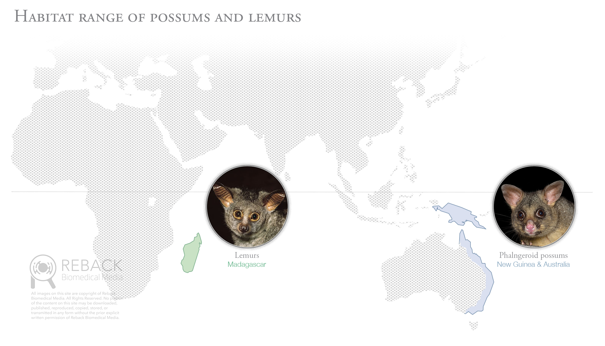
The primary goal of this project was to digitally reconstruct the masticatory apparatus of a single species of phalangeroid possum, Trichosurus vulpecula. The reconstruction serves as a visual communication tool that improves the ability of the evolutionary biologist to make meaningful comparisons between the phalangeroid possums of New Guinea and Australia to lemurs of Madagascar. Lemurs, a diverse group of primitive primates, are important in the study of primate origins.

The secondary goal of this project was to document the digital reconstruction workflow to serve as an instructive guide for those interested in making their own 3D representations of mammalian masticatory apparatuses.

Why study Phalangeroids?

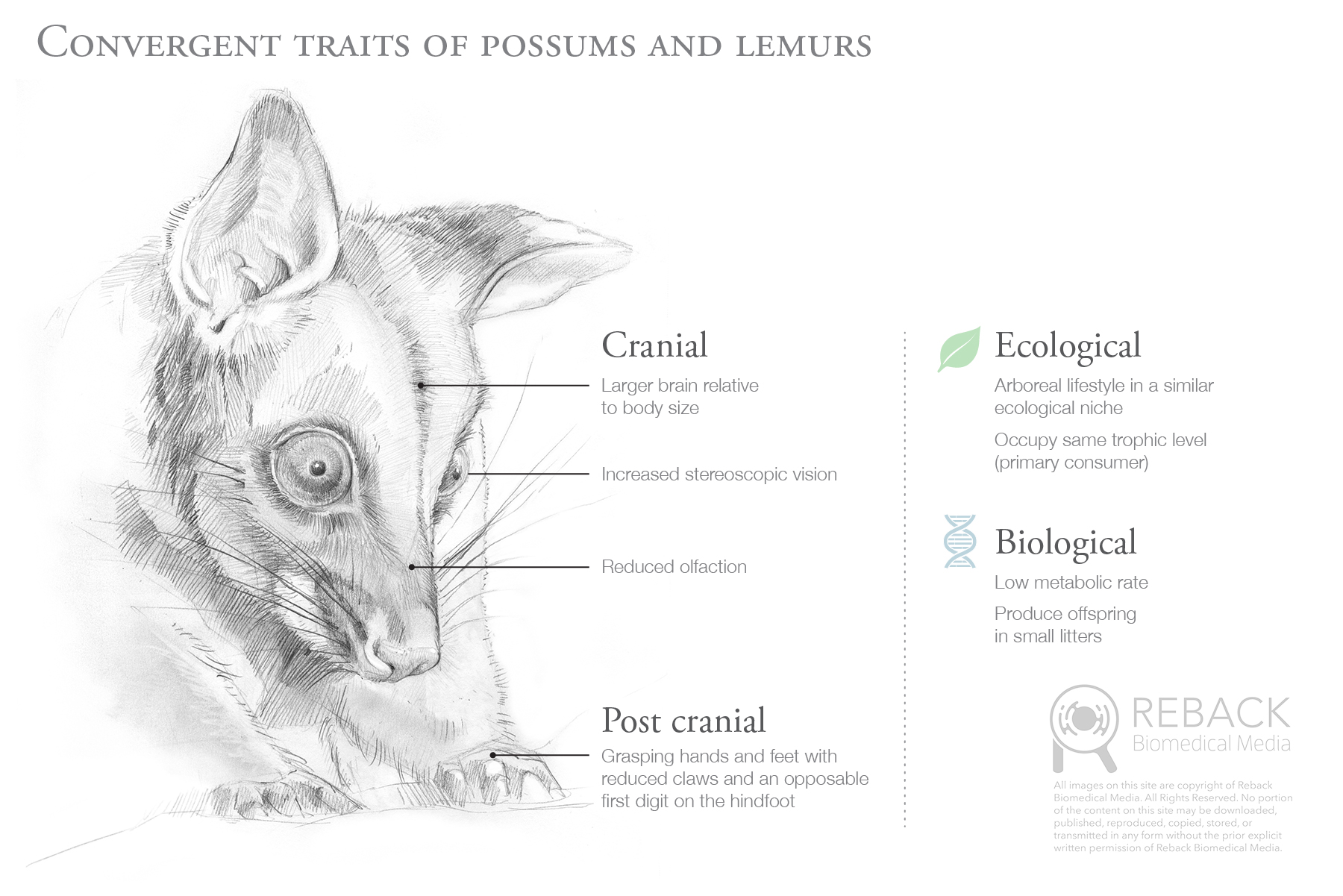
The phalangeroid possums of New Guinea and Australia are a group of mammals possessing many morphological and biological traits that are highly similar to the lemurs of Madagascar. Lemurs and phalangeroids are an example of a phenomenon known as convergent evolution: they are not closely related, yet developed similar traits as a result of having to adapt to similar ecological niches. 
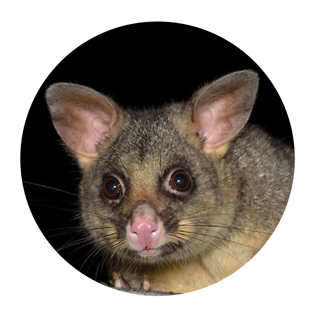
PHALANGEROID POSSOM

Trichosurus vulpecula
A marsupial mammal of the family Phalangeridae native to the island of New Guinea and east coast of Australia.
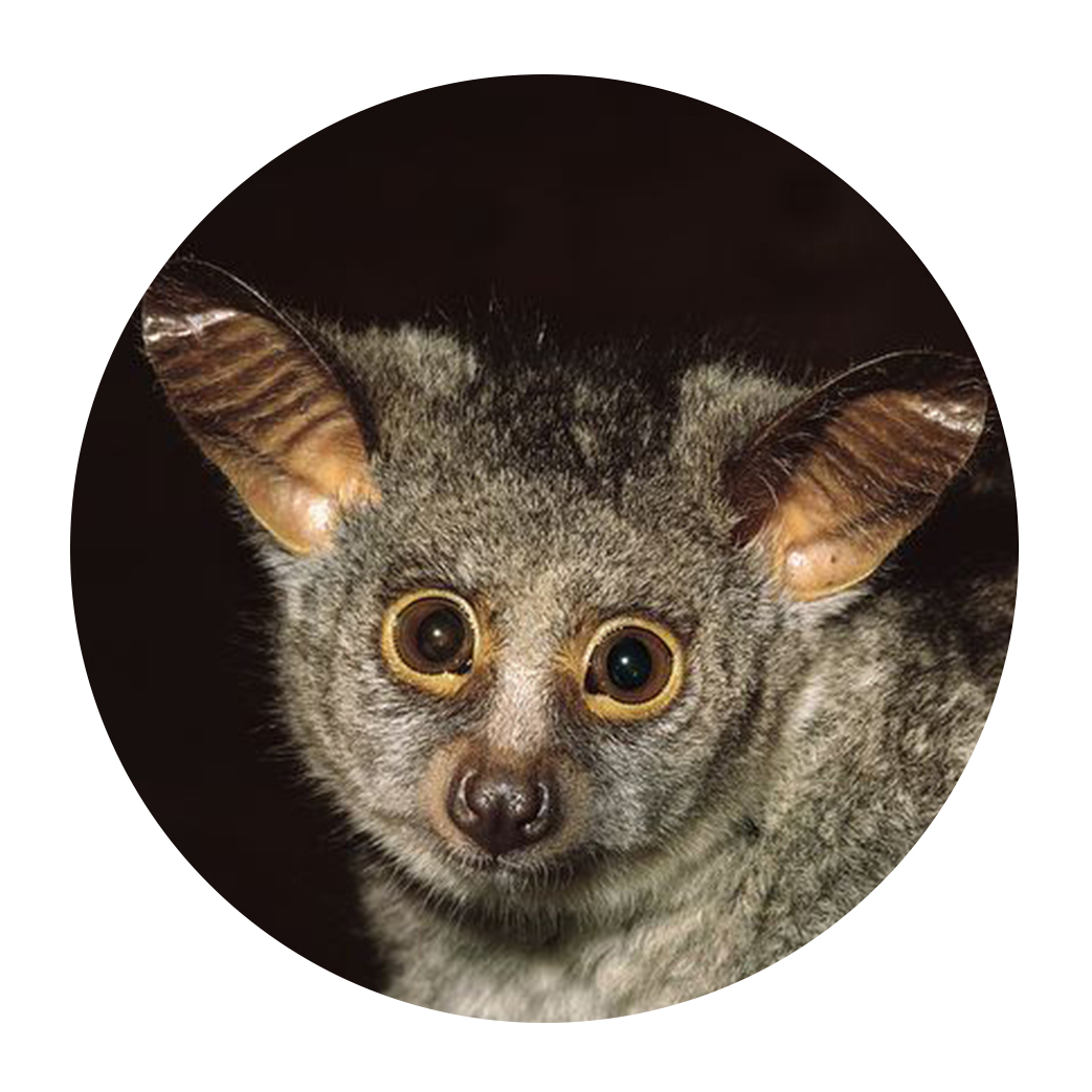
LEMUR

Otolemur crassicaudatus
A placental mammal of the order Primate native to the island of Madagascar.

Background

isolated habitats

Possums and lemurs evolved in separate and isolated regions of the world, however, the environmental conditions in each geographic region are highly similar.

Shared traits

The similarity of ecological niches inhabited by possums and lemurs influenced these two unrelated groups of mammals to develop highly similar traits.

Independent evolution

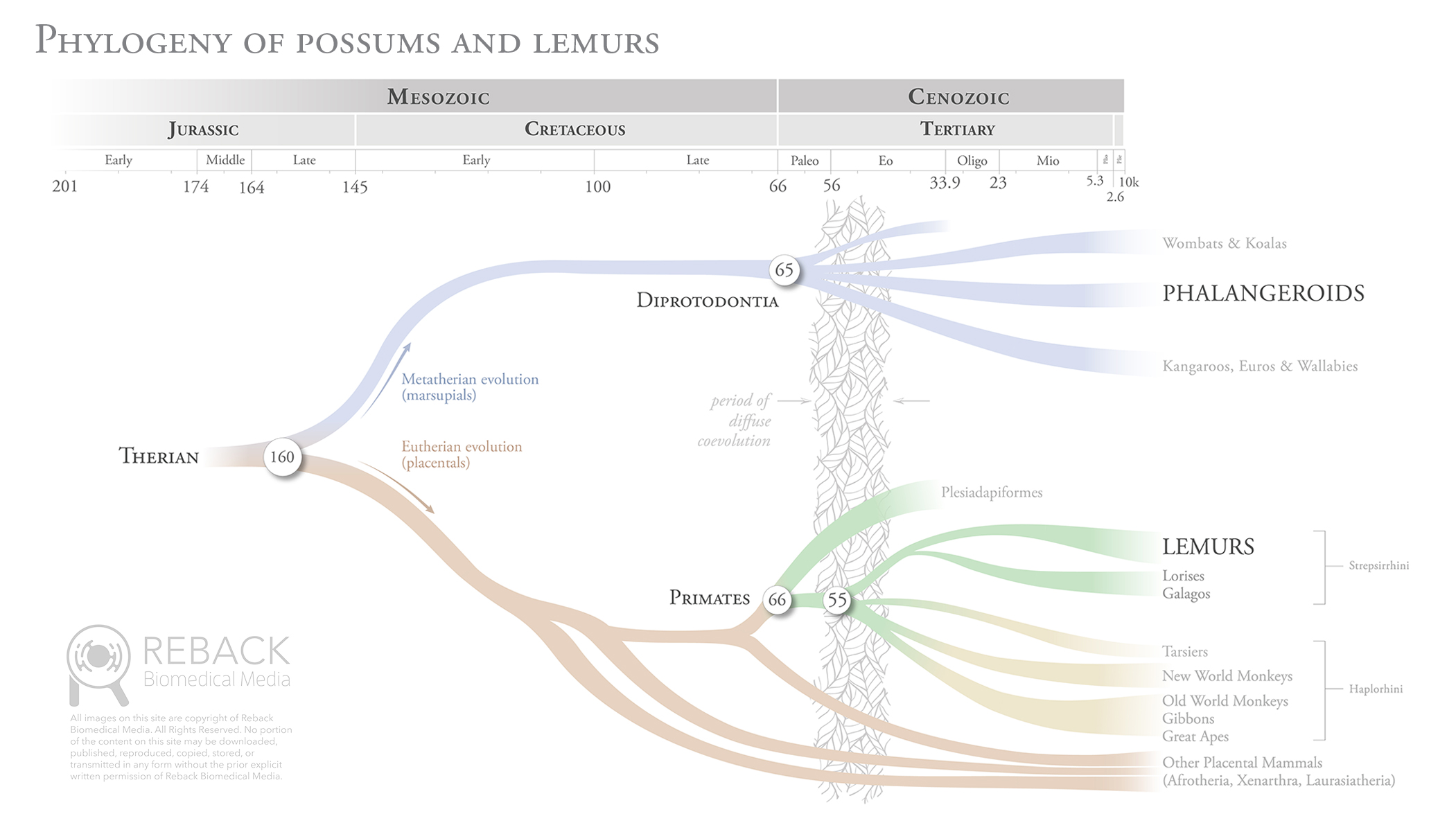
The last common ancestor to both possums and lemurs was a lizard-like mammal called a therian who lived approximately 160 mya. In fact, the diversity of all mammalian life can be traced back to this single predecessor.

reconSTruction process

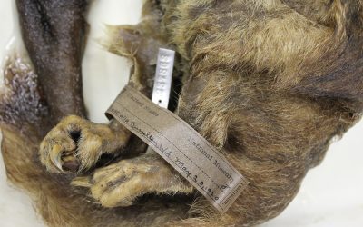
Dissection of several preserved Trichosurus vulpecula specimens took place at the Smithsonian National Museum of Natural History Museum Support Center outside of Washington, D.C. Data was gathered on muscle mass, fiber direction, and areas of origin and insertion. 

results

anatomical landmarks

Illustration was used to document the anatomical landmarks of the cranium and mandible. Observations gathered during dissection helped determine locations of muscle origin (red) and insertion (blue).

3D reconstruction

Hard tissue reconstruction relied upon segmenting data acquired from a micro-CT scan of a Trichosurus skull. Muscles were created from scratch using digital sculpting software. The mass of each muscle was matched to the average weight taken from
dissected specimens.

3D Animation

A turntable animation was created to give the audience an overall sense of spatial relationships in the masticatory apparatus. An exploded view animation demonstrates the layering of the muscles. Deeper structures, such as the pterygoids, are visualized by seeing through the mandible.

Play Video

A new TOOL FOR VISUAL COMMUNICATION

Articulated 3D PRINTED MODEL

The final output of this project was an articulated 3D printed model at 200% scale. Neodymium magnets were used to allow the audience to take apart and reassemble the muscle layers as well as separate the mandible from the skull. An axis was created in the temporomandibular joint allowing the mandible to be articulated when the muscles are removed. Several prototypes were created to design and test various physical features that made the model easy and fun to interact with.

acknowledgements

I would like to thank my thesis advisor, Jennifer Fairman, MA, CMI, FAMI, and preceptor Jonathan M.G. Perry, MSc, PhD, for their intellectual, strategic, and emotional support throughout the project. I would also like to thank my classmates, particularly Sarah A. Chen, MA, MD, for her encouragement to step away now and then and have a beer (or two). My family and friends are a constant source of support in anything I do, so a special thanks goes out to them as well.

Click the button below to download a free pdf copy of the full thesis, including tips and techniques on how to create digital 3D reconstructions of your own.

Reback Biomedical Media | 608.852.3233 | rebackbiomed@gmail.com

All content © Reback Biomedical Media, 2015-2021, unless otherwise noted. All rights reserved.

JHU – Reno Epidural Anastomosis

Embolization of the Reno epidural anastomosis for nutcracker syndrome

PROJECT

Pathology 2D animation

AUDIENCE

Candidates for surgical treatment of Nutcracker syndromen

DESCRIPTION

Renal nutcracker syndrome is a rare disorder where blood flow coming out of the kidney is restricted. The restriction is caused by compression of the left renal vein by the superior mesenteric artery. In moderate to severe cases, patients can experience blood in the urine (hematuria) and chronic abdominal pain, pelvic pain, flank pain, fatigue, or a combination thereof.

The purpose of this animation is to educate patients on the anatomy of nutcracker syndrome and provide and overview of several surgical treatments. One such treatment involves placing a stent within the left renal vein at a site beneath the superior mesenteric artery to prevent compression. Another treatment involves embolization of the left gonadal and/or renal epidural anastomosis to raise pressure within the left renal vein and prevent collapse.

Reback Biomedical Media | 608.852.3233 | rebackbiomed@gmail.com

All content © Reback Biomedical Media, 2015-2021, unless otherwise noted. All rights reserved.

JHU – Biological Filtration

Aerobic biological filtration for freshwater aquariums

PROJECT

Biological illustration

AUDIENCE

Aquarium technicians and home enthusiasts

DESCRIPTION

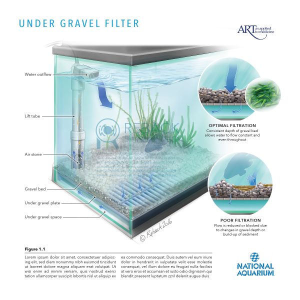
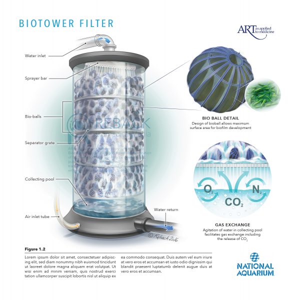
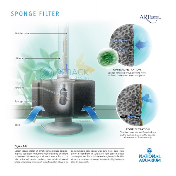
Ammonia (NH3) is a waste product excreted by fish and the decomposition of uneaten food. NH3 is toxic to fish and must be continuously filtered out of a closed system like an aquarium. Fortunately, microorganisms called nitrifying bacteria can consume NH3 as their source of energy, eventually converting it to a non-toxic substance called nitrate (NO3).

This set of illustrations depicts three different designs for aerobic biological filtration of nitrogenous waste in aquariums. Water passes through a high surface area medium (ie: gravel, bio ball, or sponge) that permits colonization of nitrifying bacteria. Oxygen is diffused into the water by either agitation (biotower filter) or pumping room air through an air stone (under gravel or sponge filter).

Details

INSETS

For more information on the nitrogen cycle in aquariums,
check out the online resources listed below:

Florida Department of Agriculture and Consumer Services:
Aquarium water quality: Nitrogen cycle

The aquarium co-op:
The easy guide to the nitrogen cycle for aquariums

Fishlore:
Aquarium nitrogen cycle – new tank syndrome


Reback Biomedical Media | 608.852.3233 | rebackbiomed@gmail.com

All content © Reback Biomedical Media, 2015-2021, unless otherwise noted. All rights reserved.
JHU – Surgical Instruments

3D modeling of surgical instruments

PROJECT

Cinema 4D three dimensional modeling and photorealistic rendering

AUDIENCE

General medical audience

DESCRIPTION

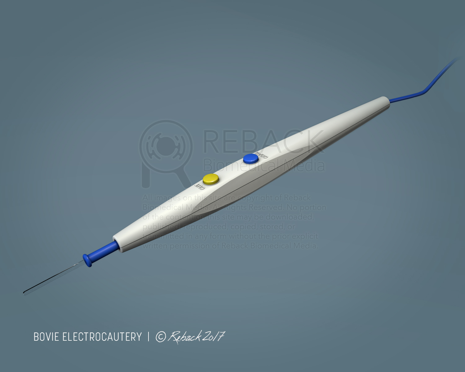
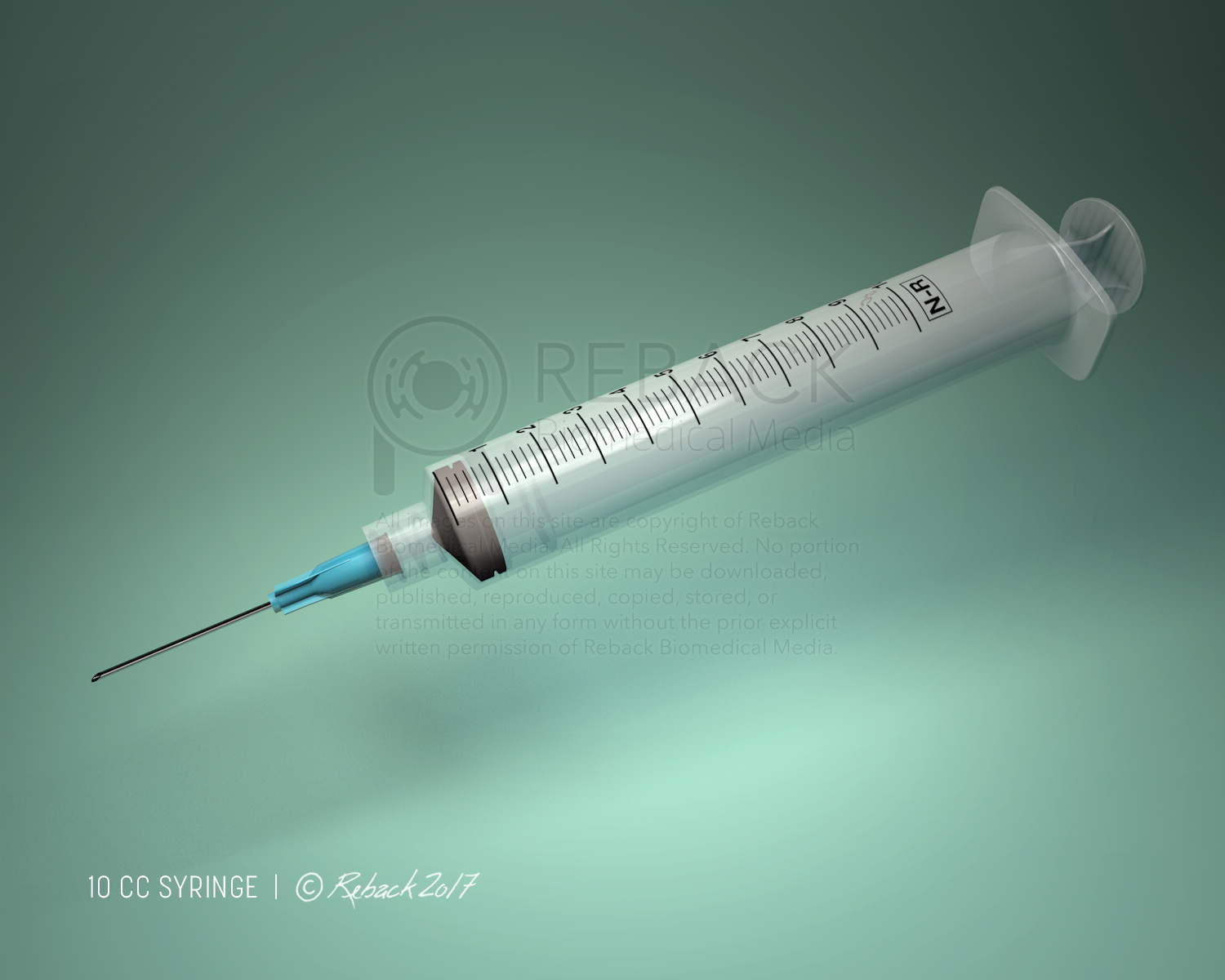
The purpose of this project was to develop basic 3D modeling capabilities in Cinema 4D for hard surface objects. A small variety of ubiquitous surgical instruments were chosen as subjects for the assignment. Importance was placed on dimensional accuracy, mechanical details, touch points, as well as color, material, texture and graphics.

Instruments were placed in a simulated studio environment with a two-light set up. Photorealistic renderings were created using the physical renderer with ambient occlusion and an HDRI studio rig to increase the perception of realistic reflections.

Reback Biomedical Media | 608.852.3233 | rebackbiomed@gmail.com

All content © Reback Biomedical Media, 2015-2021, unless otherwise noted. All rights reserved.
AtriCure – Ablation

SURGICAL ANATOMY FOR EPICARDIAL ABLATION OF
CARDIAC ARRHYTHMIA

CLIENT

AtriCure (in partnership with Merge Creative)

AUDIENCE

AtriCure device sales representatives, electrophysiologists, and cardiothoracic surgeons

DESCRIPTION

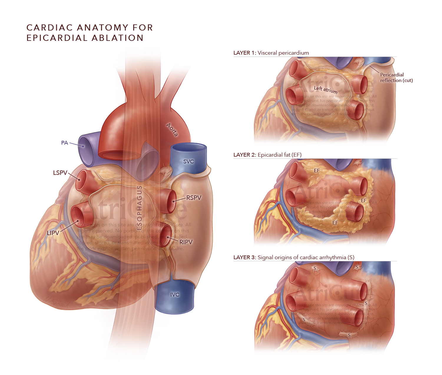
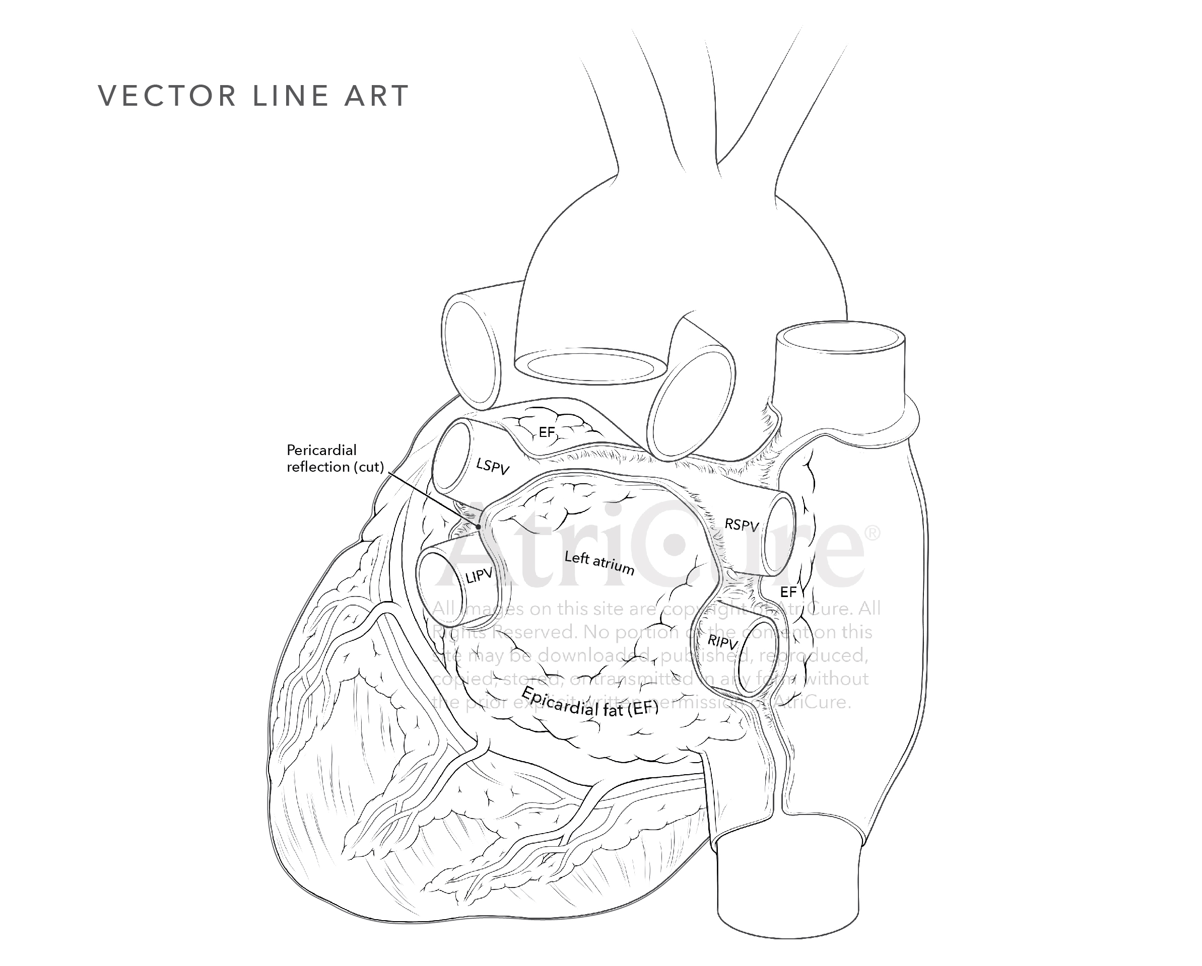
AtriCure designs and manufactures innovative technologies for treatment of atrial fibrillation and related conditions. Epicardial ablation involves approaching the exterior surface of the left ventricle through percutaneous insertion of a catheter into the pericardial space from the distal end of the sternum. 

AtriCure contacted Reback Biomedical Media to diagrammatically illustrate tissue layers surrounding the left ventricle in a posterior view. The purpose of the illustration is to facilitate communication between AtriCure device sales reps and clinicians specializing in an epicardial approach for treatment of atrial fibrillation.

PROJECT ATTRIBUTES

Timeline: 4 weeks 
Style: digital color and vector line art
Quantity: single multi-layered illustration
Scientific complexity: high

Reback Biomedical Media | 608.852.3233 | rebackbiomed@gmail.com

All content © Reback Biomedical Media, 2015-2021, unless otherwise noted. All rights reserved.
Sigvaris – Lymphatic anatomy

Lymphatic Anatomy
and Pathology

CLIENT

Sigvaris Group

AUDIENCE

Adults with impairment of lower extremity lymphatics

DESCRIPTION

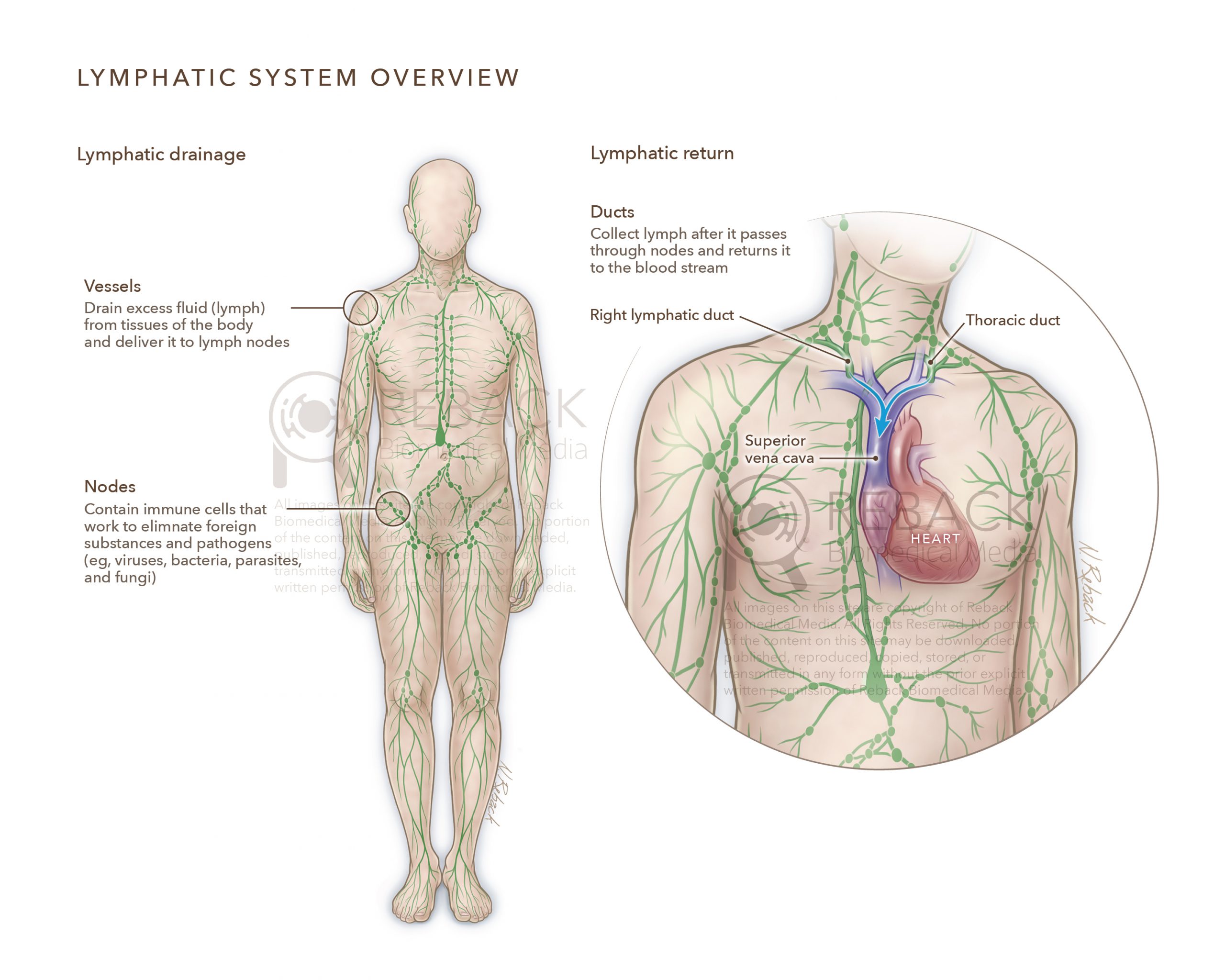
The lymphatic system consists of vessels and nodes. The vessels drain excess fluid (lymph) from the interstitial spaces of our body’s tissues then return it to the blood stream. While en route, the lymph passes through nodes where it comes into contact with immune cells (lymphocytes). The lymphocytes work to clear pathogens and other unwanted substances from the lymph.

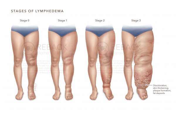
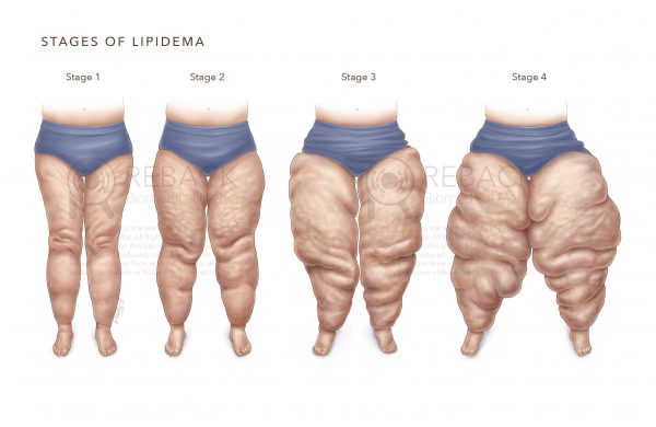
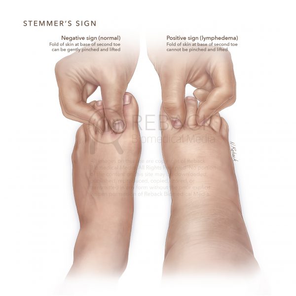
The purpose of this project is to 1) educate patients on basic lymphatic system anatomy, 2) clarify staging of lymphedema and lipidema, and 3) demonstrate a diagnostic test for lower extremity lymphedema (Stemmer’s sign).

PROJECT ATTRIBUTES

Timeline: 6 weeks
Style: full color
Quantity: 1 anatomical and 3 dermatological
Scientific complexity: medium

Reback Biomedical Media | 608.852.3233 | rebackbiomed@gmail.com

All content © Reback Biomedical Media, 2015-2021, unless otherwise noted. All rights reserved.
Sigvaris – Vein Pathology

VEIN ANATOMY AND PATHOLOGY

CLIENT

Sigvaris Group

AUDIENCE

Adults age 50 yrs and older with lower limb venous insufficiency and associated pathology

DESCRIPTION

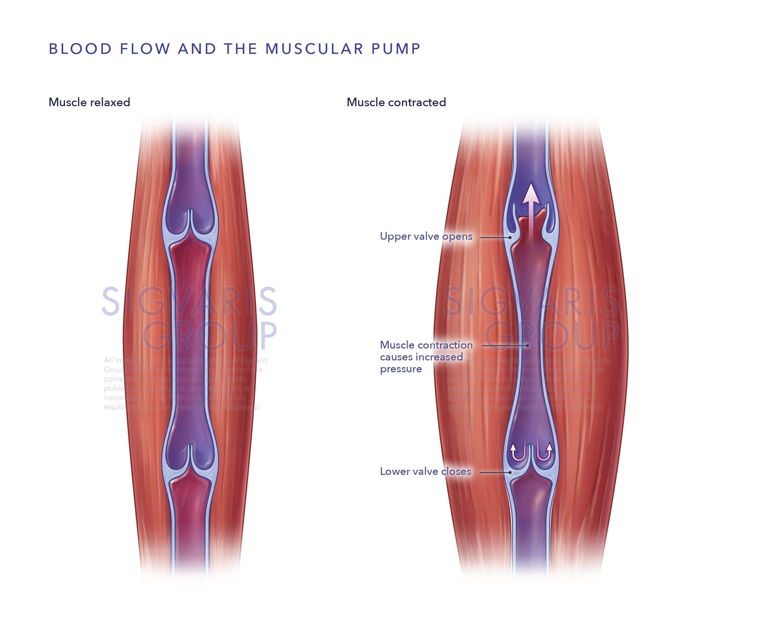
The Swiss owned company Sigvaris Group is a global provider of medical compression garments that help restore venous function and relieve associated conditions.

Sigvaris Group contacted Reback Biomedical Media to produce several anatomical and pathological illustrations for their patient web portal. The purpose of the illustrations is to 1) educate patients on normal vs abnormal venous function and 2) assist patients with identification of associated lower extremity dermatological pathology.

PROJECT ATTRIBUTES

Timeline: 8 weeks + revisions
Style: full color
Quantity: 4 anatomical and 6 dermatological illustrations
Scientific complexity: medium

Reback Biomedical Media | 608.852.3233 | rebackbiomed@gmail.com

All content © Reback Biomedical Media, 2015-2021, unless otherwise noted. All rights reserved.
Prelude DX – DCIS

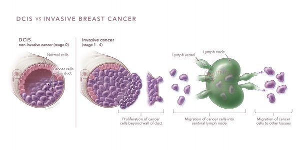
FEMALE BREAST ANATOMY AND DUCTAL CARCINOMA IN SITU (DCIS)

CLIENT

Prelude DX (in partnership with Media Directions)

AUDIENCE

Female patients with non-invasive
breast cancer (DCIS)

DESCRIPTION

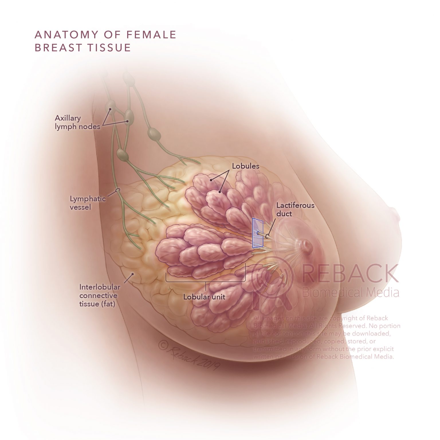
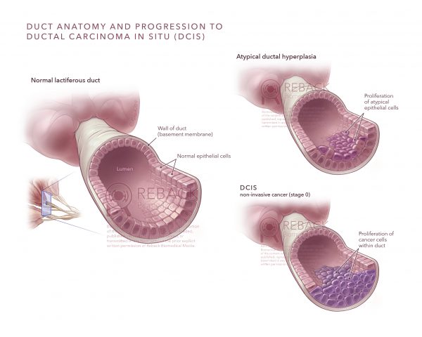
Prelude DX provides laboratory tests for patients with non-invasive breast cancer (DCIS) that help predict the overall benefit of radiation therapy when compared to other treatment modalities including surgery or active surveillance.

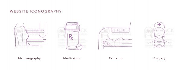
Illustrations and iconography were created for use on the Prelude DX website and in printed patient education materials. The purpose of the illustrations was to 1) explain normal anatomy of female breast tissue, 2) depict how DCIS affects a lactiferous duct at the histological level, and 3) contextualize DCIS in relation to invasive breast cancer.

PROJECT ATTRIBUTES

Timeline: 6-8 weeks + revisions
Style: digital color illustration and vector line iconography
Quantity: 5 illustrations and 4 icons
Scientific complexity: medium

Reback Biomedical Media | 608.852.3233 | rebackbiomed@gmail.com

All content © Reback Biomedical Media, 2015-2021, unless otherwise noted. All rights reserved.