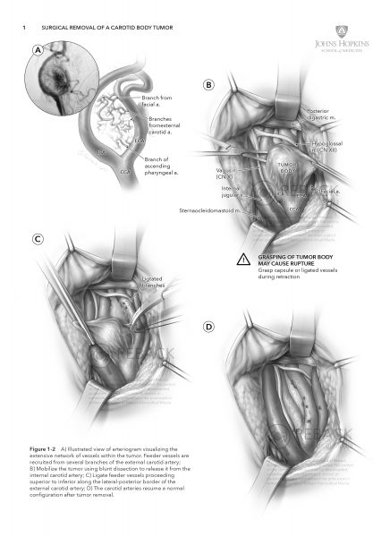
JHU – Carotid Body Tumor

Surgical Removal of
a Carotid Body Tumor

PROJECT

Surgical illustration 

AUDIENCE

Vascular and head/neck surgeons

DESCRIPTION

A carotid body tumor (also known as a chemodectoma or paraganglioma) is a rare, highly vascular mass that forms within the carotid sheath at the base of the carotid bifurcation. Tumors are often benign but may cause impairment of adjacent cranial nerves resulting in loss of associated function.

The purpose of this illustration is to teach the main steps of the surgery with special attention paid to the reaction of tissue to the instruments and other forces applied to it. The illustration was produced in collaboration with surgeons in the department of otolaryngology at the Johns Hopkins Hospital in Baltimore, Maryland. 

INSETS

layout For a surgical atlas

Reback Biomedical Media | 608.852.3233 | rebackbiomed@gmail.com

All content © Reback Biomedical Media, 2015-2021, unless otherwise noted. All rights reserved.

JAMA – Medical News

Medical news & perspectives

Articles in the Medical News & Perspectives section are focused on current events in healthcare and medical science. In-house writers chose a topic of interest and a medical illustrator is assigned to capture a particular aspect of the story in an editorial-style illustration. 

In contrast to clinically focused illustrations, those produced for Medical News are emotive and often experimental in style and technique. Although each illustration aims to be instructive in nature, the tone varies widely between them.

ARTICLE LINKS

COPYRIGHT NOTICE

All illustrations displayed on this post are copyright of the American Medical Association. All rights reserved.

Reback Biomedical Media | 608.852.3233 | rebackbiomed@gmail.com

All content © Reback Biomedical Media, 2015-2021, unless otherwise noted. All rights reserved.

JAMA – Patient Pages

PATIENT INFORMATION

JAMA Patient Information pages are a free source of one page easy-to-understand articles on prevention and management of illness or other conditions. Each article is authored by a clinician working in their specialty with whom a JAMA medical illustrator works to develop an accompanying illustration that helps explain the topic to a non-medical audience.

Access to Patient Information pages is free. No membership to the American Medical Association is required. Use the links below to view articles for each illustration shown here, or search for another topic using the JAMA Patient Information web portal.

ARTICLE LINKS

1. Patellofemoral pain
01.23.18 – JAMA

3. Vasectomy
06.19.18 – JAMA

4. Gallbladder cancer
09.25.18 – JAMA

6. Focal radiation therapy for cancer
01.10.19 – JAMA Oncology

8. Sigmoid volvulus
06.25.19 – JAMA

11. What is Mohs surgery?
05.06.20 – JAMA Dermatology

13. What are ear tubes?
10.15.20 – JAMA Otolaryngology

14. Bladder cancer
11.17.20 – JAMA

15. Urinary diversion
TBD – JAMA

COPYRIGHT NOTICE

All illustrations displayed on this post are copyright of the American Medical Association. All rights reserved.

Reback Biomedical Media | 608.852.3233 | rebackbiomed@gmail.com

All content © Reback Biomedical Media, 2015-2021, unless otherwise noted. All rights reserved.
JAMA – Difficult Intubation

WILL THIS PATIENT BE DIFFICULT
TO INTUBATE - THE RATIONAL
CLINICAL EXAMINATION

PUBLICATION DATE

02.05.2019

DESCRIPTION

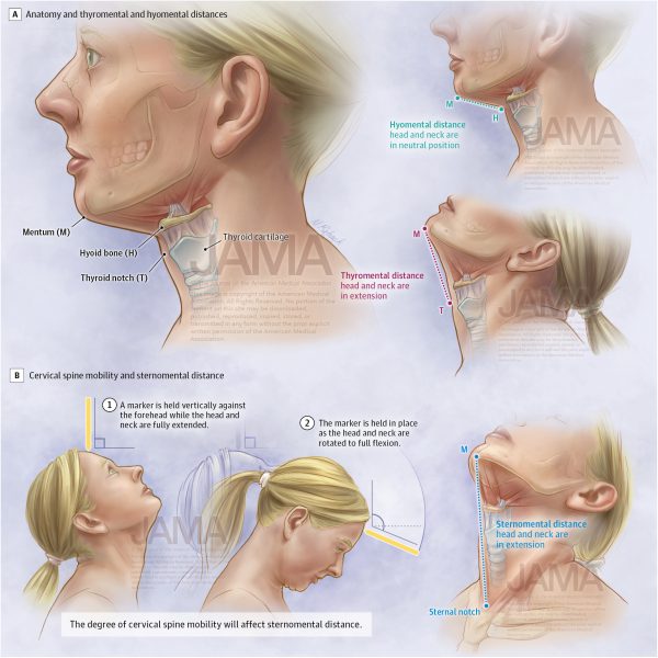
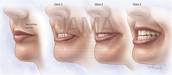
Endotracheal intubation (ETI) is often employed during major surgical procedures and for respiratory support in patients who are critically ill. Not all patients can be intubated easily, however, and failure of advanced recognition of difficult intubation can result in a catastrophic scenario.

The purpose of this illustration is to demonstrate several anatomic features and bedside tests that can be used to assess difficult intubation prior to general anesthesia or in emergency situations. This set of illustrations is unique in that it combines the most common strategies for airway assessment within a single easy-to-understand composition.

ARTICLE LINK

AMA membership required for full article access.

COPYRIGHT NOTICE

All illustrations displayed on this post are copyright of the American Medical Association. All rights reserved.

Upper Lip Bite Test

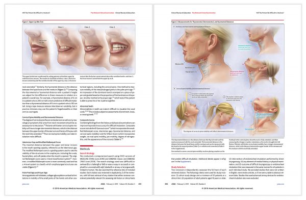
Figures in context

Reback Biomedical Media | 608.852.3233 | rebackbiomed@gmail.com

All content © Reback Biomedical Media, 2015-2021, unless otherwise noted. All rights reserved.

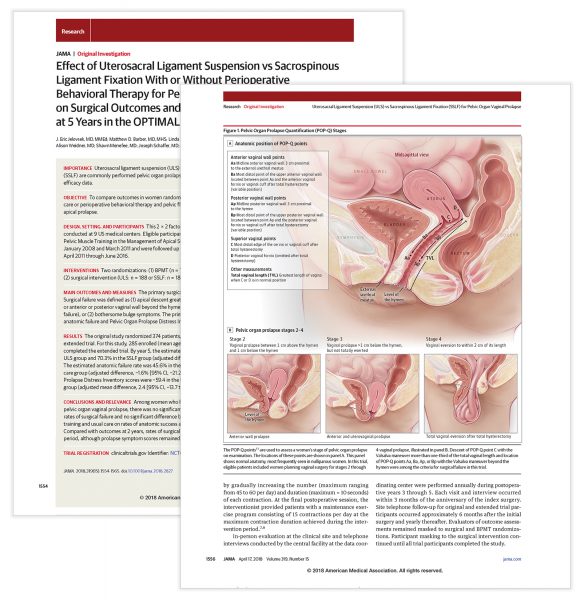
JAMA – Pelvic Organ Prolapse

Pelvic Organ prolapse Quantification -
An original investigation

PUBLICATION DATE

04.17.2018

DESCRIPTION

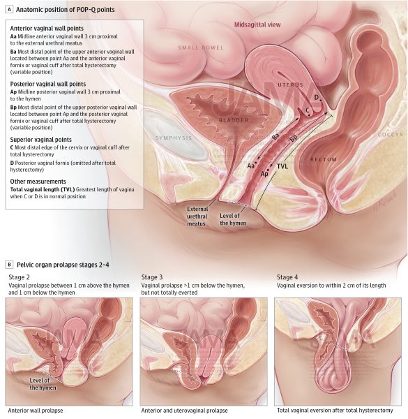
Female pelvic organ prolapse (POP) involves the downward descent of pelvic and/or lower abdominal organs resulting in protrusion of the vagina, uterus, or both. The condition is becoming more common given the aging population in the United States.

The purpose of this illustration, using normal anatomy of a nulliparous female, is to demonstrate the precise location of anatomic points used to assess POP during examination. The illustration is unique in that it is the first to unequivocally document these points and provide precise examples POP stages.

ARTICLE LINK

AMA membership required for full article access.

COPYRIGHT NOTICE

All illustrations displayed on this post are copyright of the American Medical Association. All rights reserved.

INSETS

FIgure in Context

Reback Biomedical Media | 608.852.3233 | rebackbiomed@gmail.com

All content © Reback Biomedical Media, 2015-2021, unless otherwise noted. All rights reserved.

JAMA – OSA

Diagnosis and Management of Obstructive Sleep Apnea -
A review

PUBLICATION DATE

04.14.2020

DESCRIPTION

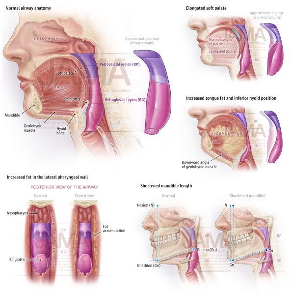
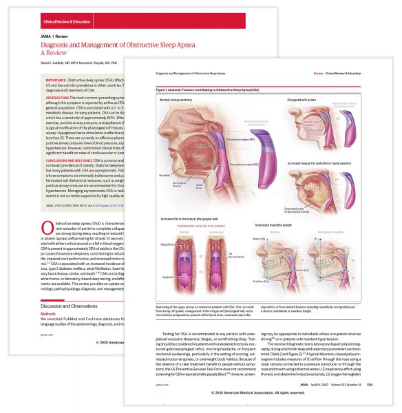
Obstructive sleep apnea (OSA) is characterized by recurrent episodes of partial or complete collapse of the upper airway during sleep. This results in the interruption of the sleep cycle and has negative consequences for quality of life. OSA is a relatively common condition, but its prevalence is increasing with rising rates in obesity.

The purpose of the illustration is to educate a broad clinical audience on anatomic variations in the upper airway that cause restriction of airflow and contribute to OSA. The illustration is innovative through the inclusion of conceptualized three-dimensional volumes alongside mid-sagittal anatomical views to demonstrate how each variation restricts airflow.

ARTICLE LINK

AMA membership required for full article access.

COPYRIGHT NOTICE

All illustrations displayed on this post are copyright of the American Medical Association. All rights reserved.

INSETS

Figure in context

Reback Biomedical Media | 608.852.3233 | rebackbiomed@gmail.com

All content © Reback Biomedical Media, 2015-2021, unless otherwise noted. All rights reserved.

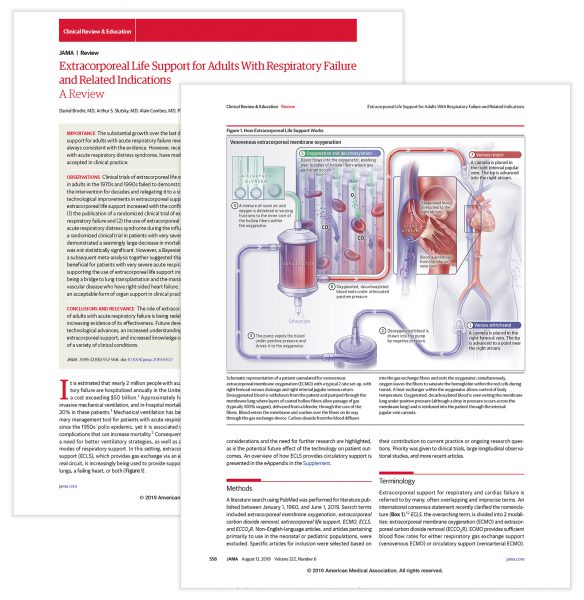
JAMA – ECMO

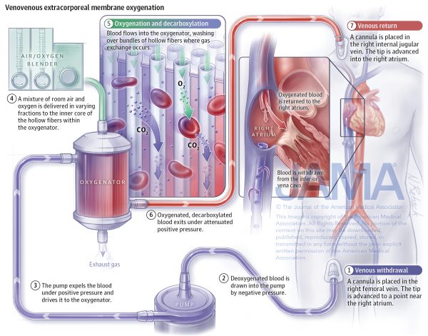
Extracorporeal membrane oxygenation - A review

PUBLICATION DATE

08.13.2019

DESCRIPTION

Extracorporeal membrane oxygenation (ECMO) is a life support system often employed in place of mechanical ventilation. It supports the work of the heart and lungs during a period of critical illness and is most commonly used for conditions such as acute respiratory distress syndrome. Recently it has played a small but important role treating severe cases of COVID-19. The purpose of this illustration is to teach a broad clinical audience how ECMO performs decarboxylation and reoxygenation in a common two-site cannulation set up.

ARTICLE LINK

AMA membership required for full article access.

COPYRIGHT NOTICE

All illustrations displayed on this post are copyright of the American Medical Association. All rights reserved.

INSETS

FIgure in Context

Reback Biomedical Media | 608.852.3233 | rebackbiomed@gmail.com

All content © Reback Biomedical Media, 2015-2021, unless otherwise noted. All rights reserved.